
Description
CHAPTER TWO: COORDINATION IN MAMMALS
Complete Study Notes & Revision Guide
📚 What This Document Covers
This comprehensive chapter breakdown covers everything you need to master coordination in mammals — from the fundamental concepts of stimuli and response to the intricate workings of the nervous system, the structure and function of neurones, the brain and spinal cord, sense organs, and the effects of drugs on nervous coordination.
🔬 SECTION 1: FOUNDATIONS OF COORDINATION IN ANIMALS
Understanding Coordination
Coordination is the linking together of different parts or organs in the body so that they work at the appropriate time and required rate. Without coordination, body activities would be disorganized and the body would fail to function properly.
The Three Requirements of Coordination
Detect changes in the environment
Transmit information to appropriate body parts
React to changes in a way that ensures survival
Stimuli and Responses
Stimuli = changes in the environment to which organisms respond
Irritability/Sensitivity = the ability to sense, interpret, and respond to stimuli
Response = a behavioral, physiological, or muscular activity initiated by a stimulus
Type of Stimulus Description Examples
External stimuli From the surrounding environment Wind, cold, heat, light, odor, vibration, pressure, pain, water, gravity
Internal stimuli From within the organism Sense of body position, glucose level, blood pressure
Why Coordination Matters — The Evolutionary Perspective
Predator avoidance: Rapid reflex responses prevent injury
Food acquisition: Coordinated movement allows hunting and foraging
Homeostasis: Maintains stable internal conditions
Reproduction: Coordinated behaviors are essential
🔑 Key Concept: The nervous system is the fast coordination system (electrical impulses, milliseconds), while the endocrine system is the slow coordination system (hormones, minutes to hours).
🧠 SECTION 2: NERVOUS COORDINATION IN HUMANS
Overview of the Nervous System
Division Components Function
Central Nervous System (CNS) Brain and spinal cord Integration, processing, decision-making
Peripheral Nervous System (PNS) Nerve fibres branching from CNS Communication between CNS and body
Homeostasis = the body’s ability to maintain stable internal conditions through feedback mechanisms.
Components of Nervous Coordination
Component Definition Examples
Stimulus Change in environment Touch, pain, temperature, light
Receptors Specialized cells that detect changes Eye, ear, nose, skin, tongue
Coordinator Receives and interprets messages Brain and spinal cord
Effectors Receive motor impulses and bring about response Muscles and glands
Response Change shown by organism Pulling hand from hot object
Feedback Animal decides what to do in response —
The Coordination Pathway
text
Stimulus → Receptor → Coordinator (CNS) → Effector → Response
↑ |
└──────────── Feedback ────────────────┘
🔑 Key Concept: The coordinator uses messages called nerve impulses — electrical signals transmitted along neurones.
🔬 SECTION 3: STRUCTURE OF NEURONES
What Are Neurones?
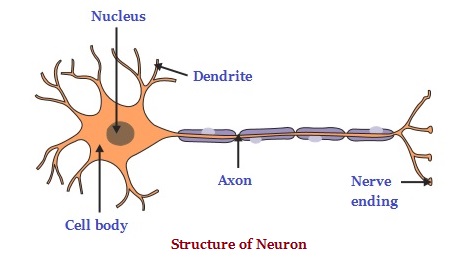
Neurones are the conducting cells of the nervous system — the basic structural and functional units found between receptors and effectors.
Distinguishing properties:
Highly responsive to stimuli
Capable of conducting nerve impulses rapidly over long distances
General Structure of a Neurone
Part Description Function
Cell body Contains nucleus; angular/oval/star-shaped Processes nerve impulses it receives
Dendrites One or more extensions from cell body Receive signals and direct them to cell body
Axon Long, stem-like part Sends signals to other cells
Axoplasm Space inside axon with charged ions Conducts electrical impulses
Myelin sheath Fatty layer covering some axons Protects, insulates, facilitates rapid transmission
Neurilemma Cellular layer inside myelin sheath Nourishes and insulates the axon
Nodes of Ranvier Periodic gaps in myelin sheath Facilitate rapid transmission by allowing “leaping”
Axon terminal Terminal end of axon Transmits signals to next neurone or effector
Why the Myelin Sheath Matters — Saltatory Conduction
Unmyelinated axons: Impulse travels continuously — relatively slow
Myelinated axons: Impulse “leaps” from node to node — saltatory conduction (from Latin saltare = to leap)
Advantages: Much faster and more energy-efficient (fewer ion movements = less ATP used)
Clinical note: Multiple sclerosis (MS) — myelin sheath damaged → saltatory conduction fails → muscle weakness, coordination problems
Types of Neurones
Feature Sensory Neurone Motor Neurone Interneurone
Other name Afferent neurone Efferent neurone Relay/intermediate neurone
Structure Long dendron, short axon Short dendrites, long axon Short dendrites, short/long axon
Cell body location Outside CNS Inside CNS Inside CNS
Function Receptor → CNS CNS → Effector Connects sensory to motor
Direction of impulse Toward CNS Away from CNS Within CNS
Adaptations of a Neurone to Its Function
Myelin sheath — protection, insulation, rapid transmission
Schwann cells — secrete myelin sheath
Many dendrites — large surface area for receiving impulses
Elongated axons — carry information over long distances
Tiny branches at end — facilitate receiving impulses from other neurones
High metabolic rate — requires glucose and oxygen for ion gradients
Long lifespan — most formed before birth, must last a lifetime
🔗 SECTION 4: THE SYNAPSE
Definition
A synapse is the gap at the point where the axon terminals of one neurone are in association with the dendrite or cell body of another neurone, or an effector organ. The gap is called the synaptic cleft.
Mechanism of Synaptic Transmission
Impulse arrives at axon terminal of presynaptic neurone
Calcium ions (Ca²⁺) enter → trigger exocytosis of synaptic vesicles
Neurotransmitter (e.g., acetylcholine) released into synaptic cleft
Neurotransmitter binds to receptors on postsynaptic membrane
Ion channels open → Na⁺ enters → depolarization → new impulse generated
Neurotransmitter broken down (e.g., by acetylcholinesterase) or taken back up → synapse reset
Why Synapses Matter
Unidirectional transmission: Impulses travel only from presynaptic to postsynaptic → one-way flow
Integration: A single neurone may receive thousands of inputs and “decide” whether to fire
Modulation: Site of learning, memory, and drug action
Key Neurotransmitters
Neurotransmitter Function Location
Acetylcholine Muscle contraction, parasympathetic NS Neuromuscular junction, autonomic ganglia
Dopamine Reward, motivation, motor control Brain (substantia nigra, VTA)
Serotonin Mood, sleep, appetite Brain (raphe nuclei)
Noradrenaline Fight-or-flight response Sympathetic nervous system
GABA Inhibitory neurotransmitter Brain
Glutamate Excitatory neurotransmitter Brain
Drug Connections
Cocaine blocks dopamine reuptake → dopamine accumulates → prolonged reward → addiction
SSRIs block serotonin reuptake → mood improvement
Organophosphate pesticides inhibit acetylcholinesterase → acetylcholine accumulates → continuous muscle contraction → paralysis and death
🧠 SECTION 5: CENTRAL NERVOUS SYSTEM (CNS)
Overview
The CNS consists primarily of the brain and spinal cord. It processes received information, coordinates, and influences activities of all body parts.
The Brain — Protection
Encased in skull (cranium)
Meninges — system of membranes
Cerebrospinal fluid — cushions, provides nutrients, removes waste
The Three Main Parts of the Brain
(a) Forebrain
Composed of: olfactory lobes, cerebrum, hypothalamus, pituitary gland
Cerebrum:
Largest part; divided into right and left hemispheres
Connected by corpus callosum
Outer portion: grey matter (cell bodies); inner portion: white matter (axons)
Folds (gyri and sulci) increase surface area
Functions: Voluntary activities, speech, reasoning, memory, decision-making, behavior, vision, imagination
Localization of function:
Lobe Function
Frontal lobe Motor control, planning, decision-making, personality
Parietal lobe Somatosensory processing (touch, temperature, pain)
Temporal lobe Hearing, language comprehension, memory
Occipital lobe Vision
Hypothalamus:
Homeostasis: Regulates temperature, fluid balance, metabolism, hormones
Involuntary activities: Appetite, sleep, thirst
Link between nervous and endocrine systems: Controls pituitary gland
Thermoregulation centre and osmoregulation centre
Pituitary gland:
“Master gland” — controls activity of most other hormone-secreting glands
Anterior pituitary: Secretes TSH, ACTH, FSH, LH, GH, Prolactin, MSH
Posterior pituitary: Stores and releases ADH and oxytocin
(b) Midbrain
Smallest part of the brain
Connects hindbrain with forebrain
Functions: Conveys information, controls involuntary activities and reflex movements of head/neck/eyes, visual and auditory reflexes
(c) Hindbrain
Composed of: cerebellum and medulla oblongata
Cerebellum:
Cauliflower-shaped structure
Functions: Body movements, balance, posture; receives impulses from skeletal muscles, tendons, inner ear
Clinical note: Damage → ataxia (loss of coordination, unsteady gait)
Medulla oblongata:
Most posterior part; connects to spinal cord
Controls involuntary and reflex activities: Breathing, heartbeat, digestion, blood vessel dilation/constriction, swallowing, sneezing, vomiting, secretion of juices, temperature regulation
Clinical note: Damage → death (controls breathing and heartbeat)
Cranial Nerves
Pairs of nerves connecting brain to head, neck, and trunk:
Olfactory nerves — smell
Optic nerves — vision
Vestibulocochlear nerves — hearing and balance
Vagus nerve — mixed motor and sensory (heart, gut, respiratory tract)
Oculomotor nerve — eye movement, pupil constriction
Spinal Cord
Structure:
Runs from head to end of trunk; encased in meninges
Lies in canal of vertebral column
Cross-section: Two identical halves enclosing spinal canal (filled with cerebrospinal fluid)
Inner region: Grey matter (cell bodies)
Outer region: White matter (myelinated axons)
Spinal nerves: Dorsal root (sensory) and ventral root (motor)
Functions:
Coordinating centre — controls involuntary actions (reflex actions)
Nerve impulse conduction pathway — between brain and rest of body
Reflex centre — fast, automatic responses
Reflex actions controlled by spinal cord:
Coughing, sneezing, blinking
Removing hand or leg when touching hot or sharp object
Clinical note: Spinal cord injury → loss of function below injury level (paralysis, loss of sensation)
Comparison of Grey Matter and White Matter
Feature Grey Matter White Matter
Colour Grey White
Composition Cell bodies of neurones Myelinated axons
Location in spinal cord Inner region Outer region
Location in brain Outer region (cortex) Inner region
Function Processing, integration Conduction of impulses
✅ QUICK REVISION CHECKLIST
□ I can define coordination and explain why animals need it
□ I can describe the coordination pathway (stimulus → receptor → coordinator → effector → response)
□ I can name the three types of neurones and their functions
□ I can label the structure of a neurone
□ I can explain saltatory conduction and why myelin matters
□ I can describe the mechanism of synaptic transmission
□ I can name key neurotransmitters and their functions
□ I can describe the three main parts of the brain and their functions
□ I can explain the structure and function of the spinal cord
□ I can compare grey matter and white matter
□ I can describe the reflex arc and its components
□ I can distinguish between simple and conditioned reflexes
□ I can describe the structure of the skin and its receptors
□ I can explain the mechanism of hearing
□ I can explain the mechanism of body balance
□ I can describe the structure of the eye and its functions
□ I can explain accommodation (near vs. distant objects)
□ I can describe common eye defects and their corrections
□ I can describe the nose (smell) and tongue (taste) mechanisms
□ I can categorize drugs and describe their effects
□ I can explain how drugs affect the synapse
□ I can describe preventive measures against drug abuse
MITIHANI POPOTE — Quality educational resources wherever you are.


Reviews
There are no reviews yet.